
 EN
EN



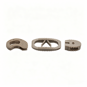
Facing spinal surgery can feel overwhelming. If your surgeon has recommended a lumbar fusion, you have likely heard them mention using a lumbar cage for lumbar fusion surgery.
Understanding exactly what this device is, how it works inside your body, and what to expect during your recovery can significantly demystify the process and help you approach your surgery with confidence.
What is a Lumbar Cage?
To understand a lumbar cage, it helps to look at the anatomy of your lower back. Your spine is made up of individual bones called vertebrae, which are stacked on top of each other. In between these bones sit intervertebral discs—rubbery cushions that absorb shock and allow your spine to bend and twist.
When a disc degenerates due to age, wear-and-tear, or injury, it can collapse. This narrowing of the space between vertebrae can cause severe back pain or pinch nearby nerves, leading to shooting leg pain (sciatica), numbness, or weakness.
A lumbar cage for lumbar fusion surgery is a small, hollow, ring-like medical device inserted directly into the space where your damaged disc used to be. Its primary job is to act as a permanent spacer, propping the two vertebrae back up to their normal, healthy height and relieving pressure on compressed nerves.
The Patient Experience: A Clinical Journey to Fusion
To understand how a lumbar cage functions in real life, consider the typical clinical journey of a patient suffering from severe degenerative disc disease.
Case Study: Regaining Mobility After Years of Pain
The Patient: A 52-year-old high school teacher had been struggling with chronic lower back pain and progressive right leg numbness for over three years. Conservative treatments—including physical therapy, epidural steroid injections, and anti-inflammatory medications—no longer provided relief, severely limiting her ability to stand and teach.
The Problem: An MRI revealed severe disc collapse at the L4-L5 level, causing the bone to rub against bone and severely pinching the exiting nerve root.
The Surgical Solution: The surgeon performed a minimally invasive Transforaminal Lumbar Interbody Fusion (TLIF). First, the damaged, collapsed disc material was carefully removed. Next, a titanium lumbar cage packed with bone graft material was inserted into the empty disc space.
The Outcome: The cage immediately restored the collapsed disc height, creating wide-open channels for the pinched nerves. Over the next six months, the patient’s natural bone grew through and around the hollow center of the cage, fusing the L4 and L5 vertebrae into one solid, stable bone block. Today, she is back in the classroom, completely free of leg numbness and managing only mild, standard postoperative stiffness.
Materials and Mechanics: What Is Inside Your Back?
Lumbar cages are engineered to be incredibly strong, biocompatible (meaning your body will not reject them), and structurally supportive. Today, surgeons primarily use two advanced materials for these cages:
-
PEEK (Polyetheretherketone): PEEK is a high-grade, medical-plastic polymer.
-
The Advantage: Its structural flexibility is very similar to natural human bone, which prevents the cage from putting too much stress on the surrounding vertebrae. Because PEEK is radiolucent (transparent on X-rays), it allows your surgeon to easily track your bone healing progress during postoperative checkups.
-
-
Titanium Alloy: Titanium is a time-tested biocompatible metal, and modern titanium cages often feature highly advanced, porous surfaces.
-
The Advantage: Human bone cells love titanium. The microscopic, rough texture of a 3D-printed porous titanium cage mimics natural bone structure, actively encouraging your own bone cells to grow directly onto and into the implant—a process called osseointegration.
-
Why is the Cage Hollow?
The hollow center of a lumbar cage for lumbar fusion surgery is critical. Before insertion, the surgeon packs this empty space with a bone graft (either small pieces of your own bone, donated bone, or a synthetic bone-growing protein). The cage holds the spine steady immediately after surgery, while the bone graft inside does the long-term work of growing the bridge that locks the two vertebrae together permanently.
Biomechanical Consensus and Long-Term Success
The use of lumbar cages is considered the gold standard in modern spinal fusion because it solves two major mechanical problems simultaneously:
-
Immediate Primary Stability: Before the cage was invented, surgeons relied solely on bone grafts alone to maintain disc height. These grafts could easily slip, crack, or compress under the weight of the body. A lumbar cage provides immediate, rigid mechanical support the moment it is placed, keeping your spine aligned while you heal.
-
Maximizing Fusion Rates: According to extensive data published by organizations like the North American Spine Society (NASS) and the AO Foundation, utilizing an interbody lumbar cage significantly increases successful fusion rates compared to older fusion techniques that left the disc space empty. By holding the bones perfectly still and maintaining tension on the surrounding ligaments, the cage creates the perfect biological environment for solid bone growth.
Summary for Patients
If your surgical plan includes a lumbar cage for lumbar fusion surgery, remember that this tiny device is engineered to protect you. It restores your natural spinal alignment, takes the pressure off your painful nerves, and serves as the protective scaffolding your body needs to heal and stabilize your lower back for the long haul. Always talk openly with your spine specialist about the specific material and surgical approach that is safest and most effective for your unique anatomy.